Read in more detail in our publication:
- Shkedrov, C., Florshaim, Y., Ness, G., Gandman, A., Sagi., Y. High Sensitivity RF Spectroscopy of a Strongly-Interacting Fermi Gas, Phys. Rev. Lett. 121, 093402 (2018).
In short
We investigate of an ultracold Fermi gas in the BCS-BEC crossover using a novel RF spectroscopy technique we have developed.
We report on two main results:
- For the first time, we were able to probe the rf lineshape tail all the way to the energy scale characterizing the short-range interactions. In comparison to previous studies, we extend the range of frequencies experimentally accessible by more than an order of magnitude. At this extended range, we observe a universal power-law scaling of the rf lineshape over more than two decades. We demonstrate this universality for different interaction strengths in the vicinity of the Feshbach resonance. Therefore, our experimental findings corroborate the contact-like potential description of interactions between ultracold atoms up to the van der Waals scale.
- We measure the Feshbach molecule dissociation spectrum in an extended range of magnetic fields. From this data we extract the binding energy of the Feshbach molecule with very good precision (2.5%). This data clearly and systematically deviates from theoretical calculations using the most recent calibration of the Feshbach resonance parameters. We use a two-coupled channels model of the resonance to extract a new calibration of the Feshbach resonance parameters. With this calibration, both the two-channel and single channel model fit the binding energy data very well.
Both of these observations were made possible owing to unprecedented sensitivity of our new rf spectroscopy method. Standard rf spectroscopy uses absorption imaging to count the number of atoms out-coupled by the rf pulse. Our new technique uses fluorescence imaging instead, with a sophisticated scheme that transfers only the out-coupled atoms into a magneto-optical trap. This enables us to detect signals of only few atoms, which paves the way to detect the faint signals comprising the data of the two observations.
The New RF spectroscopy technique
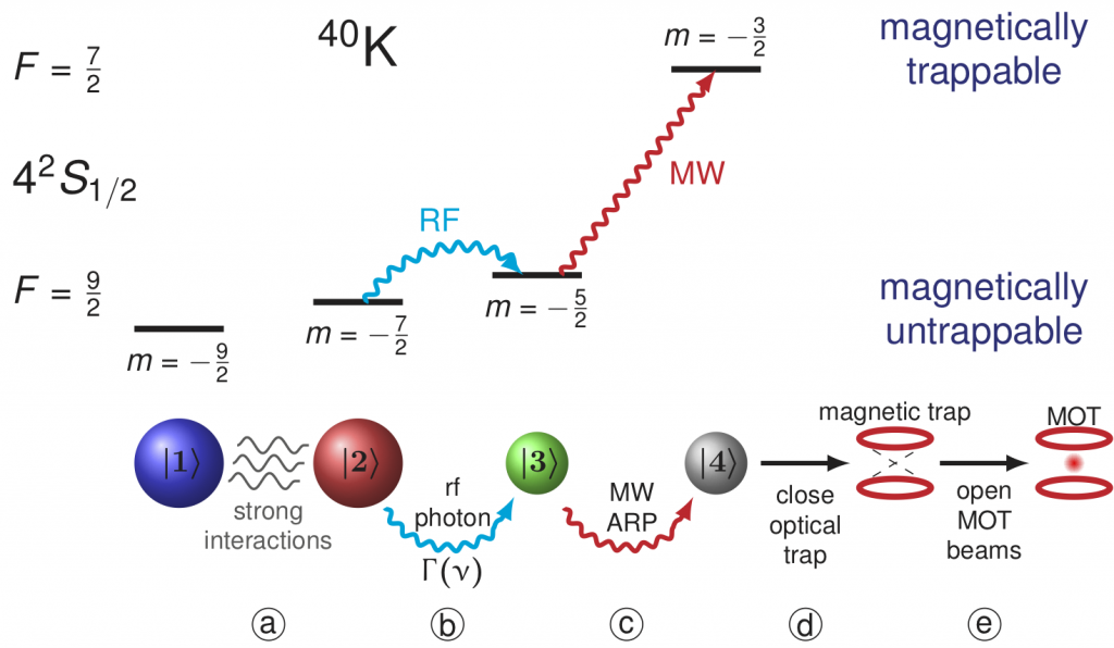
The new rf spectroscopy method is depicted in the following figure:

We prepare a quantum degenerate gas of 40K atoms in a balanced mixture of the two lowest Zeeman sublevels, denoted by |1> and |2> (a). Their energy is split by a homogeneous magnetic field B. Rf radiation at a frequency ν transfers a small fraction of the atoms at state |2> to a third Zeeman sublevel |3>, which is initially unoccupied (b). The number of atoms out-coupled by the rf pulse is recorded, and the result of this measurement is the rf transition rate Γ(ν). Many important physical observables can be extracted from Γ(ν), including pair size, superfluid gap, quasi-particle dispersion, etc. In standard rf spectroscopy, the number of the out-coupled atoms is measured by absorption imaging. Since the photon absorption cross-section is relatively small, detecting very weak signals in the rf lineshape tail is challenging. We overcame this difficulty by using fluorescence imaging instead. With fluorescence imaging, even a single atom can be reliably detected if held for a sufficient period of time, as was demonstrated with a magneto-optical trap (MOT). A MOT is a common tool in atomic physics which uses the forces exerted by a continuous scattering of photons to cool and trap atoms. To be used with rf spectroscopy, the main challenge is to capture in the MOT only the atoms out-coupled by the rf pulse, which may be a tiny fraction of the whole cloud.
Our approach relies on the difference in energy and magnetic dipole moments of the various energy levels. Specifically, all three states |1>,|2>, and |3> have negative magnetic dipole moments (“high-field-seekers”) and are therefore magnetically untrappable. After the rf pulse is applied, we use a narrow microwave (MW) sweep to selectively transfer only the population at state |3> to a state |4> in the other hyperfine manifold (c), which has a positive magnetic dipole moment and is therefore magnetically trappable (“low-field-seeker”). We then turn on a quadrupole magnetic field which magnetically traps the atoms in state |4> and expels all the rest (d). Finally, we turn on the MOT laser beams and record the MOT fluorescence signal with a sensitive CMOS camera (e). The detection sensitivity can in principle reach a single atom (in our apparatus it is limited to 2-5 atoms due to background scattering from surrounding optical surfaces).
